Mesothelioma: Staging and Treatment
encontrar mi
What is staging for cancer?
Staging is the process of learning how much cancer is in your body and where it is. For mesothelioma, x-ray, CT scan,echocardiogram, PET scan, MRI, and blood tests like Fibulin-3 and soluble mesothelin-related peptides (SMRP’s) may be done to help stage it. Your providers need to know about your cancer and your health so that they can plan the best treatment for you.
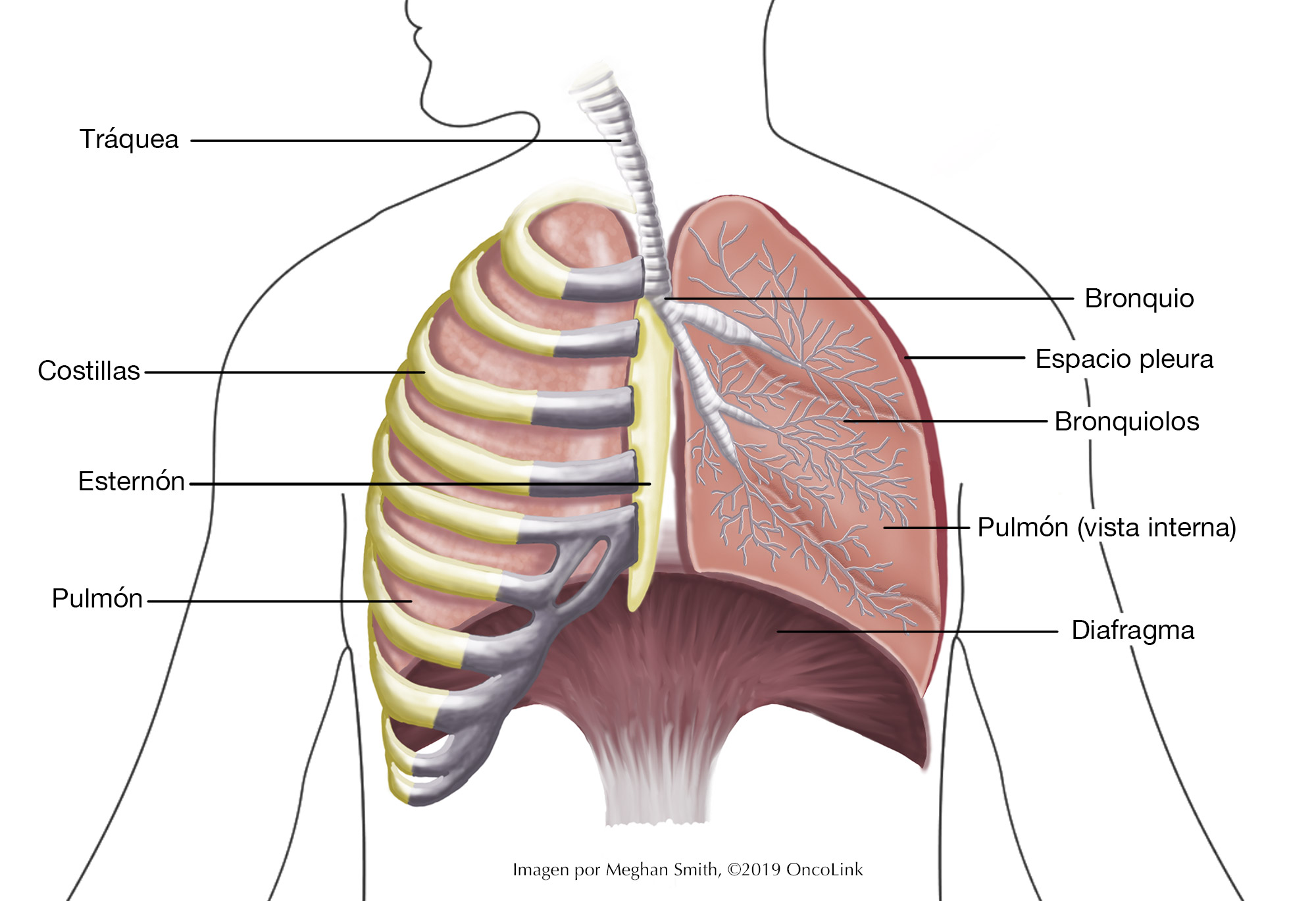
Staging looks at the size of the tumor, where it is, and if it has spread to other organs. Currently, there is only a staging system for pleural mesothelioma.
A staging system called the “TNM” system,” as described by the American Joint Committee on Cancer, helps to guide the staging of mesothelioma. It has three parts:
- T-describes the size/location/extent of the "primary" tumor in the lung.
- N-describes if the cancer has spread to the lymph nodes.
- M-describes if the cancer has spread to other organs (metastases).
How is mesothelioma staged?
Your healthcare provider will use the results of the tests you had to figure out your TNM result and combine these to get a stage from I to IV. Mesotheliomas can be classified in different ways based on the part of the body they start in. Pleural (lung) mesothelioma is the most common. The staging system for pleural mesothelioma is very complex. Below is a summary. Talk to your provider about the stage of your cancer.
Stage IA (T1, N0, M0): The cancer is on one side of the chest in the pleura lining. It may also be in the diaphragm (the muscle between the lungs), the mediastinum (space between the lungs), or covering the pleura lining of the lung (T1). The cancer is not in any of the lymph nodes (N0), or any other body parts (M0).
Stage IB (T2, N0, M0): The cancer is on one side of the chest in the pleura lining. It is also in the pleura lining the diaphragm, mediastinum, and the lung. The cancer is also inside the diaphragm or the lung (T2). The cancer is not in any of the lymph nodes (N0), or any other body parts (M0) OR (T3, N0, M0): The cancer has grown to nearby areas and may be able to be taken out with surgery (T3). The cancer is on one side of the chest in the pleura lining and, in the pleura, lining the diaphragm, mediastinum, and lung on the same side. The cancer has grown into at least one:
- Endothoracic fascia (first layer of chest wall).
- Fatty tissue in the mediastinum.
- One area in the deeper chest wall layers.
- Pericardium covering (outer layer of the heart).
- The cancer is not in any of the lymph nodes (N0), or any other body parts (M0).
Stage II (T1 or T2, N1, M0): The cancer is on one side of the chest in the pleura lining (T1). The cancer is also inside the diaphragm or the lung (T2). Mesothelioma is in nearby lymph nodes on the same side as the other tumor (N1). The cancer is not in any other body parts (M0).
Stage IIIA (T3, N1, M0): The cancer has grown to nearby areas and may be able to be taken out with surgery (T3). The cancer is on one side of the chest in the pleura lining and, in the pleura, lining the diaphragm, mediastinum, and lung on the same side. The cancer has grown into at least one:
- Endothoracic fascia (first layer of chest wall).
- Fatty tissue in the mediastinum.
- One area in the deeper chest wall layers.
- Pericardium covering (outer layer of the heart).
Mesothelioma is in nearby lymph nodes on the same side as the other tumor (N1). The cancer is not in any other body parts (M0).
Stage IIIB (T1-T3, N2, M0): The cancer may or may not be in the nearby areas and may be able to be taken out with surgery (T1 to T3). The cancer has spread to the lymph nodes on the other side of the body or the lymph nodes near the collarbone on either side (N2). The cancer is not in any other body parts (M0) OR (T4, Any N, M0): The cancer has grown too much and cannot be taken out with surgery (T4). The cancer is on one side of the chest in the pleura lining and, in the pleura, lining the diaphragm, mediastinum, and lung on the same side. The cancer has grown into at least one:
- More than one area in the deeper chest wall layers such as muscle or ribs.
- Through the diaphragm and peritoneum (abdomen lining).
- Any of the mediastinum organs (esophagus, thymus, trachea, blood vessels.)
- Spine.
- In the pleura on the other side of the chest.
- Pericardium lining or inside the heart.
The cancer may or may not be in the nearby lymph nodes (any N). The cancer is not in any other body parts (M0).
Stage IV (Any T, any N, M1): The cancer may or may not be in nearby organs (any T). The cancer may or may not have spread to the lymph nodes (any N). The mesothelioma has spread to organs, bones, the liver, the lung, the pleura on the other side of the body, or the peritoneum (M1).
How is mesothelioma treated?
Treatment for mesothelioma is based on the stage. Your treatment may include some or all of these:
- Surgery.
- Radiation.
- Chemotherapy.
- Palliative treatment.
- Clinical trials.
Surgery
Younger, healthy patients with early-stage malignant pleural mesothelioma may be able to have surgery that removes the mesothelial tissue around the tumor. Surgery to either remove the entire lung with the tumor (called extrapleural pneumonectomy) or lung-sparing surgery that removes only the tumor and the lining of the lung (which is called extended or radical pleurectomy), is the most common option for these patients.
Radiation
Radiation is the use of high-energy X-rays to kill cancer cells. Radiation therapy is often done after surgery to remove the entire lung to kill any remaining cancer cells. Radiation therapy after lung-sparing surgery is not routinely given. The radiation can damage the healthy lung tissue and result in side effects that outweigh any benefit of radiation. Radiation therapy is often delivered to surgical incision sites to prevent the cancer from recurring (coming back) in that area. In patients who do not undergo surgery, radiation therapy may be given to treat problem areas to relieve symptoms (called palliative radiation), like pain or trouble breathing.
Chemotherapy
Chemotherapy is the use of medications to kill cancer cells and is a common treatment for mesothelioma. Medications that are used, either alone or in combination, include cisplatin, carboplatin, doxorubicin, pemetrexed, gemcitabine, nivolumab, ipilimumab, pembrolizumab, and vinorelbine.
Palliative Treatment
Palliative therapy is used to relieve symptoms that are caused by the cancer. They do not cure the cancer. There are many options for palliative treatments including chemotherapy, radiation, surgery, stent placement, laser therapies, and removal of extra fluid from around the heart, lungs, or abdomen (belly). Some of the palliative treatments for mesothelioma are having a pleural catheter put in, paracentesis, and thoracentesis. Talk to your provider about your options for managing your symptoms.
Clinical Trials
You may be offered a clinical trial as part of your treatment plan. To find out more about current clinical trials, visit the OncoLink Clinical Trials Matching Service.
Making Treatment Decisions
Your care team will make sure you are included in choosing your treatment plan. This can be overwhelming as you may be given a few options to choose from. Take the time to meet with different providers and think about your options and what is best for you. This is a personal decision. Friends and family can help you talk through the options and the pros and cons of each, but they cannot make the decision for you. You need to be comfortable with your decision – this will help you move on to the next steps. If you ever have any questions or concerns, be sure to call your team.
You can learn more about mesothelioma at OncoLink.org.