Primary CNS Lymphoma
encontrar mi
Based on the OncoLink e-Case of the Month for September 1995 by James H. Johnson, Jr., MD of The Children's Hospital of Philadelphia
Case History
History of Present Illness
A 15-year old girl presents to the emergency room with a complaint of recurrent headache, blurry vision of 10 days duration and a 10-lb weight loss over the past two months. She was well until approximately ten days prior to admission when she developed diffuse non-throbbing headaches that were partially or completely relieved by oral acetaminophen. During the two to three days prior to admission, she had recurrent and constant headaches and had blurry vision while reading and watching television.
Past Medical/ Surgical History
Unremarkable other than tonsillectomy at age 7.
Medications/ Allergies
None reported.
Social History
The patient attends high school and lives with parents and younger brother. She has no history of tobacco, ethanol or drug abuse.
Review of Systems
As per HPI
Physical Examination
GENERAL: mildly cachectic, appears stated age
VITAL SIGNS: afebrile with vital signs within normal range for age.
SKIN: without rashes
HEENT: pupils equal, round and reactive to direct light; normal funduscopic exam with no retinal abnormalities
PULMONARY: normal breath sounds, symmetric chest expansion
CARDIOVASCULAR: normal, no murmurs, rubs or gallops
ABDOMINAL: without organomegaly, spleen tip not palpable
GENITOURINARY: normal
EXTREMITIES: no abnormalities noted
LYMPH: no palpable lymphadenopathy.
NEURO:
- alert and oriented in all spheres, normal speech and affect
- cranial nerve examination reveals a mild left VIth nerve palsy, but no nystagmus
- motor examination reveals mild left upper extremity drift, normal muscle tone and
- bulk rapid alternating movements are normal
- sensory examination is intact to soft touch, pinprick, vibration and proprioception
- gait demonstrates normal stance and arm swing with normal heel, toe and tandem gait
- cerebellar examination demonstrates normal finger-nose and heel-shin coordination
- reflexes are increased (3+) at the left biceps, patella and ankle
- negative Romberg's sign, no clonus, absent Babinski's sign
Data
LABS: Within normal range
IMAGING: An emergent cranial CT is obtained which demonstrates an abnormal density of the thalami with mild hydrocephalus, but no evidence of acute hemorrhage or mass effect. The initial CT scan with administration of IV contrast demonstrates bilateral thalamic mass lesions that enhance. There is mild hydrocephalus, edema surrounding the masses and mass effect on adjacent CNS structures.
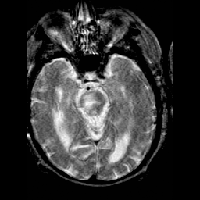
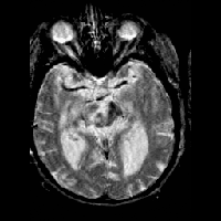
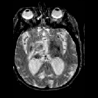
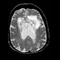
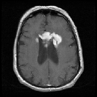
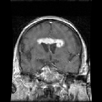
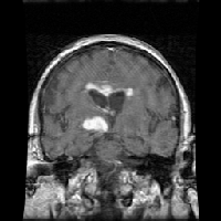
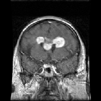
To further define the lesion, a cranial MRI with intravenous gadolinium is obtained. The MRI also demonstrates bilateral thalamic mass lesions with significant enhancement following IV administration of gadolinium. Again, hydrocephalus and mass effect are demonstrated. The dense signal characteristics are suggestive of a highly cellular tumor such as a primitive neuro-ectodermal tumor (PNET) or lymphoma.
Cranial MRI Images |
DIAGNOSTIC STUDIES: She is treated with high dose intravenous dexamethasone for relief of mass effect-related symptoms for approximately 24 hours (six mg every six hours) prior to a stereotactic biopsy.
The surgical biopsy specimen consists of tiny fragments of tissue and clot. Normal brain tissue is identified with reactive astrocytes. The tumor sample consists of mononuclear cells. Large and small lymphocytes are noted with a relatively high number of macrophages predominating the field. There are also areas of micronecrosis of lymphocytes. Mitoses are identified in a few large cells. Immunoperoxidase techniques searching for various antigens and markers produce the following findings:
Immunoperoxidase Staining Results
B cells | ++++ positive in large lymphocytes | NFP | ++++ positive in residual |
T cells | ++++ positive in small lymphocytes | PcNA | 60-80% positive |
CD 68 | ++++ positive in macrophages | HCG, CEA, AFP | all negative |
GFAP | ++++ positive in reactive astrocytes (supporting or glial cells of the central nervous system) |
The pathologic diagnosis based on the morphology and special stains is B-cell Lymphoma of the large cell type, with necrosis secondary to steroids.
Assessment and Plan
According to the histopathology of the surgical biopsy specimen, the patient's diagnosis appears to be a B-cell non-Hodgkin's lymphoma, of the large cell subtype. At this point a thorough staging evaluation is warranted for appropriate prognostic and therapeutic management. This evaluation consists of a complete CT scan of the chest and pelvis with oral and IV contrast, a bone scan, a bone marrow biopsy and aspirate, and analysis of cerebral spinal fluid.
Clinical Course
The patient's staging work-up is entirely negative. Follow-up neuro-imaging 24 hours after biopsy demonstrates a marked reduction in the mass lesions of the thalami.
She is initially treated with six months of adjuvant chemotherapy consisting of cyclophosphamide, vincristine, prednisone, doxorubicin, high-dose methotrexate (3 gm/m2) and cytosine arabinoside. Her course is complicated by severe, chemotherapy-related mucositis requiring IV fluids and morphine and also by central diabetes insipidus not requiring medical therapy. A post-chemotherapy cranial MR scan documents a very good response. An end-of-chemotherapy MRI demonstrates evidence of a surgical defect, resolved hydrocephalus and minimal residual disease.
Consolidative therapy is achieved with 45.0 Gy total brain irradiation, administered in a helmet distribution. She currently has no evidence of disease shortly after the completion of her radiation therapy.
Discussion
Malignant lymphomas are categorized into Hodgkin's disease and non-Hodgkin's lymphoma (NHL). CNS involvement by NHL may occur as a primary lesion without evidence of nodal or visceral occurrence, or as a secondary infiltration either from direct extension or distant hematogenous metastases. Additional terms in the nomenclature of primary CNS lymphoma include reticulum cell sarcoma, immunoblastic sarcoma and microglioma. Modern classifications based on immunohistochemistry have recognized lymphocytic elements and the monoclonality of populations. The spectrum of CNS lymphomas includes the spectrum of nodal lymphomas: histiocytes, plasma cells, B cells, T cells and microglial cells. Most, however, are of B cell lineage, while T cell and histiocytic lines are rare.
Historically, primary CNS-NHL constitute a small percentage(<2%) of all central nervous system tumors in adults, with a peak age of onset of 55 years. The incidence of primary CNS-NHL is slightly higher in males than females. The majority of patients at major cancer treatment centers are members of the AIDS and HIV-infected populations as well as other immunocompromised patients, including transplant recipients. In children, the majority of cases have similarly been in AIDS patients. Recently, however, there have been several reports demonstrating an increase in the number of non-AIDS adult patients with primary CNS lymphoma. Two studies, the SEER data collected by the NCI and an Italian population based study, noted that the rate of primary CNS lymphoma is on the rise in the immunocompetent population.
In addition to AIDS and HIV, primary CNS lymphoma may occur in other acquired or congenital immunosusceptible conditions such as the Wiskott-Aldrich syndrome, hyperimmunoglobulin A, and in medical immunosuppression from chronic corticosteroids or anti-transplant rejection agents such as cyclosporin. The only known infectious agent related to primary CNS lymphoma is Epstein-Barr virus exposure.
Diseases Associated with Primary CNS Lymphoma
Congenital or acquired immunodeficiencies |
Presentation
The clinical presentation of primary CNS lymphoma depends on the specific location of the infiltrating lesion or the mass effect on adjacent normal structures. In general, CNS lymphoma is an infiltrating lesion that appears diffuse or as a mass lesion with non-specific neurologic signs and symptoms. Rarely, it may appear without a mass lesion and only a monoclonal cell population in the cerebral spinal fluid is detectable. There may be headache that is often non-localized and general malaise due to increased intracranial pressure, with a slowly developing confusional state clinically similar to encephalitis. When the orbits are involved, there may be ocular pain or visual disturbances including uveocyclitis and visual field abnormalities. Specific neurologic findings may be helpful in localization to a specific region of the brain. Initial evaluation varies depending on the immunologic status of the patient.
In immunocompetent and immunosuppressed patients, recent studies in adults with primary CNS lymphoma indicate differing early diagnostic evaluations. Adults with AIDS who develop CNS lymphoma have a large incidence of orbital and meningeal involvement with dissemination to other organ systems including bone marrow. However, in the immunocompetent patient, primary CNS lymphoma appears to be limited to the neuro-axis, with rare involvement of the bone marrow and visceral organs.
Radiographic examination with CT and /or MRI of the cranium provides essential information at diagnosis as to the extent of dissemination and overall mass size of the lesion. CT scan may demonstrate an area of low signal that enhances homogeneously with IV contrast. Cranial MRI provides improved anatomical definition. Multiple imaging modalities may be used to demonstrate the lesion and enhancement with IV gadolinium. Additional sites of CNS axis dissemination include but are not limited to the orbits, brainstem, meninges and spine.
In children, primary CNS lymphoma is rare and its natural history and presentation is less clear. The majority of cases to date have been in children with AIDS and other immunodeficiencies. The limited anecdotal experience in the diagnostic evaluation of lymphoma outside the CNS axis in children appears to correlate with the adult experience.
Treatment
Historically, conventional therapy in adults involved high-dose corticosteroids with cranial or craniospinal radiation therapy. Survival was poor: generally 12-18 months in non-AIDS patients and 3-5 months in AIDS patients. Recent trials utilizing adjuvant, non-Hodgkin's style chemotherapy have shown possibly improved survival.
There may be some theoretical and practical reasons to use pre-irradiation chemotherapy. Initial chemotherapy can demonstrate sensitivity of lymphoma cells to anti-neoplastic agents including cytoreduction. Recent clinical trials of chemotherapy using HD-MTX as initial therapy followed by cranial radiotherapy have demonstrated significantly improved survival in the AIDS and non-AIDS patients. One study at Memorial Sloan-Kettering Cancer Center demonstrated a mean disease-free survival of 40 months with the longest survivor out at nine years after intravenous and intrathecal MTX.
Cranial radiation therapy has been the mainstay of treatment. Several doses and techniques have been evaluated. Early studies used doses of 40-50 Gy with improved survival, but a dose-response study has not as yet been done. Currently, most centers are utilizing a dose of 45-50 Gy combined with pre-irradiation chemotherapy with good outcome, including disease-free survival up to 54 months in one adult study. The use of brachytherapy and radiosurgery may be beneficial as palliative treatment if relapse occurs.
The role of surgery is limited to biopsy. Retrospective analysis of a large adult population who received resection versus biopsy alone showed no difference in survival.
Survival and quality of life of patients with primary CNS lymphoma can be improved with chemotherapy and cranial radiation therapy after pathologic diagnosis. Direct extrapolation of the adult experience may not be entirely applicable to children. Improved chemotherapeutic agents and regimens require additional study in both adults and children.
